|
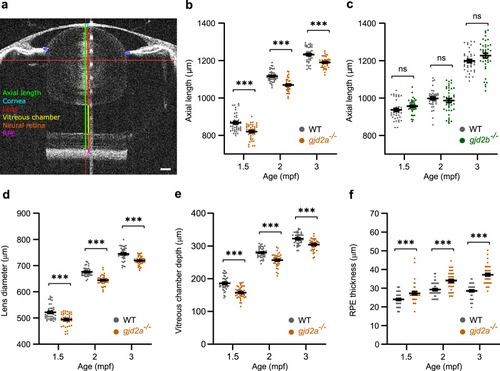
Loss of Cx35.5 (<italic>gjd2a</italic>) leads to reduced ocular dimensions.SD-OCT recordings of size-matched juvenile-to-adult zebrafish indicating the temporal dynamics of the eye. All ocular metrics were corrected for the tissue-specific refractive index. a Single B-scan image of a 3 mpf zebrafish eye. The area is defined as the axial length that spans from the apical part of the corneal epithelium to the anterior border of the RPE (green line). The RPE is represented by the hyperreflective melanin-rich band (magenta), of which the anterior part comprises a sharp-cut border, used as a posterior landmark for the axial length. The gradient refractive index of the spherical zebrafish lens was used as a correction factor to acquire this image (see “Methods”), and the brightness was enhanced for better visualization of the transparent lens. Individual compartments: cornea (light blue), lens (red), vitreous chamber (yellow), neural retina (orange). b, c Axial length of gjd2a (Cx35.5) (b) and gjd2b (Cx35.1) (c) mutant eyes of juvenile (1.5–2 mpf) and adult zebrafish (3 mpf). b The gjd2a (Cx35.5) eyes were significant reduced in axial length compared with WT at 1.5 mpf (effect size = −47 µm, p < 0.001), 2 mpf (Effect size = −48 µm, p < 0.001), and 3 mpf (effect size = −43 µm, p < 0.001). c The gjd2b (Cx35.1) mutant eyes showed no significant alteration relative to WT. d, e, f Dimensions of significantly altered ocular compartments in gjd2a (Cx35.5) mutant eyes. See Supplementary Data S1 and S2 for a full statistical report and dimensions of individual compartments. Sample size: n = 40 eyes for each genotype and time point. Error bars: SEM. Significance: ns = not significant, *p < 0.05, **p < 0.01, ***p < 0.001. Scale bar: 100 µm. Mpf months post-fertilization, SD-OCT spectral-domain optical coherence tomography, RPE retina pigmented epithelium.
|