Fig. 6
- ID
- ZDB-FIG-210514-10
- Publication
- Benson et al., 2021 - Photoactivatable metabolic warheads enable precise and safe ablation of target cells in vivo
- Other Figures
- All Figure Page
- Back to All Figure Page
|
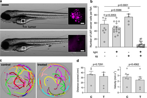
a Representative brightfield microscopy images (from five independent experiments) of zebrafish larvae grafted with U87-nlsCrimson cells before treatment (top) and after the injection of compound 15 (6 pg per embryo) and light irradiation (37 J cm−2, bottom). Insets show fluorescence microscopy images (λexc/em: 561/645 nm) of microtumors formed by U87-nlsCrimson cells, which are fully eliminated after treatment. Scale bars: 50 µm (brightfield), 20 µm (fluorescence). b Quantification of fluorescent U87-nlsCrimson cells remaining in zebrafish larvae under different treatments. Data presented as means ± SEM (n = 5 independent experiments). c Microscope images of superimposed pseudo-colored swimming tracks of control untreated zebrafish larvae and double-treated (compound 15 plus light) larvae (n = 5 per group, representative videos for each group shown as Supplementary Movies 1 and 2). d Quantification of distance and velocity of control untreated zebrafish larvae (C) and double-treated zebrafish larvae (i.e., compound 15 plus light, T) larvae. Data presented as mean values ± SEM (n = 5 independent experiments). P values were obtained from one-way ANOVA with multiple comparisons in (b) and two-tailed unpaired t tests in (d). Source data are available. |

