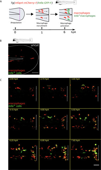
Tg(mfap4:mCherry-F/tnfa:GFP-F) reporter line allows imaging of macrophage M1-like activation at the wound in response to fin fold injury. (A) The caudal fin fold of Tg(mfap4:mCherry-F/tnfa:GFP-F) larvae were amputated at 3 dpf. Schedule of the recruitment and M1-like activation of macrophages at the wound after the fin fold injury. (B) Representative image of uncut zebrafish fin fold. Maximum projection of the overlaid fluorescence of mCherry-F (macrophages) and GFP-F (tnfa+ cells). The white line outlines the fin fold and the notochord. Scale bar: 100 μm. (C) Macrophage movements and activation states were imaged by confocal microscopy from 1.5 to 9.5 hours post amputation (hpA) using the tg(mfap4:mCherry-F/tnfa:GFP-F) line. Tail images are representative maximum projections of the overlaid fluorescences of mCherry-F (macrophages) and GFP-F (tnfa+ cells), showing M1-like activation of recruited macrophages at the wound, starting from 3 hpA and increasing up to 7.6 hpA. Dashed line indicates the wound margin. Scale bar: 100 μm.
|
