FIGURE

Figure 1

ID
ZDB-FIG-210409-1
Publication
Kayman Kürekçi et al., 2021 - Knockout of zebrafish desmin genes does not cause skeletal muscle degeneration but alters calcium flux
Other Figures
All Figure Page
Back to All Figure Page
Figure 1

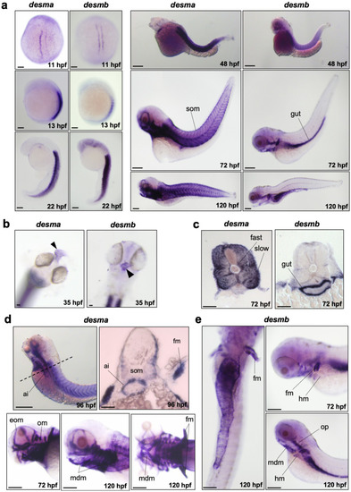
Whole mount in situ mRNA hybridisation of embryos at the indicated stages for antisense probes to desma and desmb. (a) Lateral views are anterior to top, dorsal to left for 13–22 hpf; anterior to left except 11 hpf which is a dorsal view. 48–120 hpf whole mounts are anterior to left, dorsal to top. Scale bar: 250 µm. (b) Arrows indicate heart in frontal view for left panel (desma) and ventral view for right panel (desmb) of 35 hpf embryos. (c) Transversal sections where dorsal is top. (d) Upper left panel is a lateral view at 96 hpf, scale bar: 250 µm. Dashed line represents the position of the transversal section in upper right panel where dorsal is top, scale bar: 100 µm. Lower panels are lateral views of zebrafish heads at 72 and 120 hpf treated with desma probe. Scale bar: 100 µm. (e) Left panel is a ventral view with anterior at top. Right panels are lateral views. Ai, anterior intestine; eom, extraocular muscles; fast, fast muscles; fm, pectoral fin muscles; hm, hypaxial muscles; mdm, mandibular muscles; om, opercular muscles; slow, slow muscles; som, somites. Scale bar: 250 µm.

Expression Data
Genes:
Fish:
Anatomical Terms:
Stage Range: 1-4 somites to Day 5

Expression Detail
Antibody Labeling
Phenotype Data

Phenotype Detail
Acknowledgments
This image is the copyrighted work of the attributed author or publisher, and ZFIN has permission only to display this image to its users. Additional permissions should be obtained from the applicable author or publisher of the image. Full text @ Sci. Rep.