
Fig. 5
- ID
- ZDB-FIG-210407-26
- Publication
- Fernandes et al., 2020 - Neural circuitry for stimulus selection in the zebrafish visual system
- Other Figures
- All Figure Page
- Back to All Figure Page
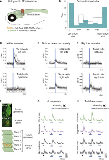
(A) Schematic of the holographic optogenetics experiment. (B) Opto-activation index. Unilateral optogenetic activation of lhx9-positive isthmic neurons (right side) leads to WTA and averaging activity in the tectum (in blue). The opto-activation index is calculated as follows: (responsive cells right tectum − responsive cells left tectum) / (responsive cells right tectum + responsive cells left tectum). Opto-index distribution for the control condition (ChR2 negative fish) is shown in gray. Distributions are significantly different (p = 0.038, two-sided Kolmogorov-Smirnov test). (C) Example of 10 cells from each tectum in a trial where the left tectum “won” (WTA). The black line shows mean response for all cells. Gray traces show individual cell activity. (D) Similar to (C) but for a trial where both tecta were equally active (averaging). (E) Similar to (C) and (D) but a trial where the right tectum “won” (WTA). (F) Activation of specific lhx9-positive isthmic neurons expressing Channelrhodopsin (ChR2; orange), combined with volumetric imaging of ipsilateral tectal responses (GCaMP6s, green). Up to five planes, including the tectum and NI region, were recorded simultaneously. (G) Photostimulation of lhx9-positive isthmic neurons. Some of the isthmic looming-evoked responses are unaffected by optogenetic stimulation (in gray), whereas others are either suppressed (magenta) or enhanced (green). Examples of averages of 10 cells are shown for each response type. Shaded areas represent SD. (H) Similar to (G) but for tectal cells. |