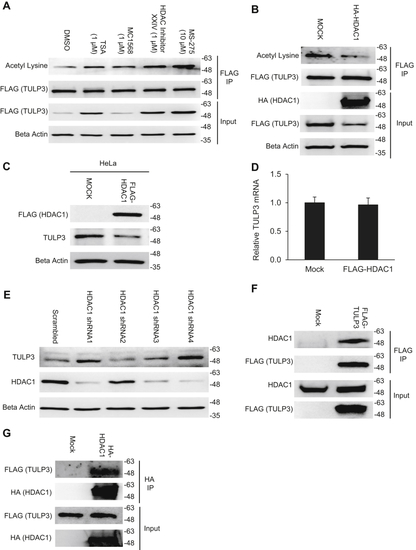
HDAC1 deacetylates TULP3 and reduces its protein levels.A, western blot showing acetylation and total protein levels of stably expressed FLAG-TULP3 in 293T cells treated with DMSO, 1 μM TSA, 1 μM MC1568, 1 μM HDAC Inhibitor XXIV, or 10 μM MS-275 for 16 h. B, acetylation and total protein levels of stably expressed FLAG-TULP3 in 293T cells transfected with empty pcDNA 3.1(+) or a plasmid encoding HA-HDAC1. Cells were harvested 48 h posttransfection. C, western blot showing endogenous levels of TULP3 in Hela cells transfected with either empty pcDNA 3.1(+) or a plasmid encoding FLAG-HDAC1. Cells were harvested 48 h posttransfection. D, mRNA levels of endogenous TULP3 corresponding to treatments in (C) analyzed by qRT-PCR; n = 4 technical replicates, Mean ± S.D. shown. E, western blot of HDAC1 and TULP3 protein levels following stable transduction of HDAC1 shRNAs into HeLa cells. Western blots showing coimmunoprecipitation of F FLAG-TULP3 with HDAC1 or G HA-HDAC1 with FLAG-TULP3 in 293T cells.
|
