
Fig. 5
- ID
- ZDB-FIG-210319-123
- Publication
- Cavanah et al., 2020 - A nontoxic fungal natural product modulates fin regeneration in zebrafish larvae upstream of FGF-WNT developmental signaling
- Other Figures
- All Figure Page
- Back to All Figure Page
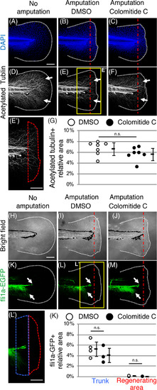
Colomitide C does not affect density of nerve and endothelial cells during larvae fin regeneration. Images of 6 dpf larvae fin without amputation (A, D). Images of 6 dpf larvae fin, amputated at 4dpf (B, C, E, F). Larvae in the middle and right column are treated with DMSO and colomitide C, respectively. DAPI (A‐C) and acetylated tubulin (D‐F) signals after whole‐mount immunostaining. Tail fin without amputation (A, D), with amputation and DMSO‐treatment (B, E, E'), and with amputation and colomitide C‐treatment (C, F) are shown. E' shows a closeup indicated in E. Red dotted area in E' show the regenerating fin. (G) Relative density of the nerve in the regenerating fin.. n.s., not significant by t‐test. Bright field images (H‐J) and fli1a‐GFP signals (K‐M) of the tail fin without amputation (H, K), with amputation and DMSO‐treatment (I, L, L'), and with amputation and colomitide C‐treatment (J, M). L' shows a closeup indicated in L. Red dotted area and blue dotted area in L' show the regenerating fin and the trunk area 100 μm from the amputation plane, respectively. (K) Relative density of endothelial cells in the trunk and regenerating fin. n.s., not significant by t‐test. White dotted lines and red dotted straight lines indicate the amputation plane, respectively. A‐F and H‐M are at the same scale. Scale bar in A, E', H and L', 100 μm |