
FIGURE
Fig. 3
Fig. 3
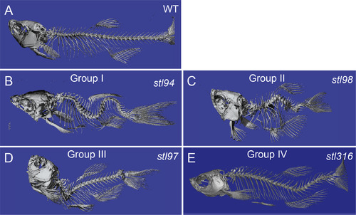
Micro-computed tomography imaging of the representative spine defect mutant classes. μCT imaging of wild type (A) and representative mutants (B–E), displaying whole-body scoliosis (stl94) (B, Group I), dwarf or shortened body plan and vertebral body defects (stl98) (C, Group II), thoracic-localized scoliosis (bhustl97) (D, Group III), and caudal-localized scoliosis locstl316 (E, Group IV). |
Expression Data
Expression Detail
Antibody Labeling
Phenotype Data
Phenotype Detail
Acknowledgments
This image is the copyrighted work of the attributed author or publisher, and
ZFIN has permission only to display this image to its users.
Additional permissions should be obtained from the applicable author or publisher of the image.
Reprinted from Developmental Biology, 471, Gray, R.S., Gonzalez, R., Ackerman, S.D., Minowa, R., Griest, J.F., Bayrak, M.N., Troutwine, B., Canter, S., Monk, K.R., Sepich, D.S., Solnica-Krezel, L., Postembryonic screen for mutations affecting spine development in zebrafish, 18-33, Copyright (2020) with permission from Elsevier. Full text @ Dev. Biol.