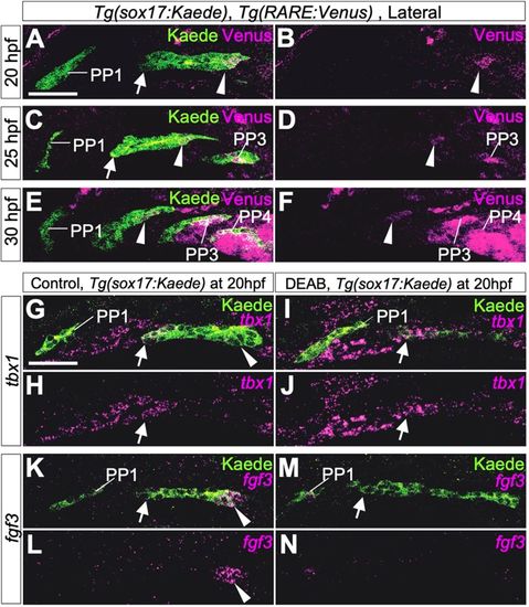
Boundaries of molecular mechanisms forming PPs between the rostral and caudal portions of PP2. (A-F) Immunohistochemistry of double transgenic embryos of Tg(sox17:Kaede) and Tg(RARE:Venus) showed specific signals of the RA reporter Venus in the caudal part of PP2 and in posterior PPs but not in the rostral edge of PP2 and PP1 endoderm at 20 hpf (A,B), 25 hpf (C,D) and 30 hpf (E,F). Three embryos were analyzed at each time point. (G-N) Expression of tbx1 (magenta in G-J) or fgf3 (magenta in K-N) in control (G,H,K,L) and DEAB-treated transgenic (I,J,M,N) embryos carrying sox17:Kaede R2 (arrows) at 20 hpf. Expression of tbx1 was not affected in DEAB-treated embryos (G,J, indicated by arrows). By contrast, RA deficiency caused by DEAB treatment resulted in loss of fgf3 expression in C2 (K-N, indicated by arrowheads). Owing to the angle at which images were taken, the size and morphology of each PP appear to be slightly different in each confocal plane, but, in these experiments, DEAB treatment did not cause major anomalies in morphology of PP1 or PP2. More than five embryos were used for each experiment.
|
