FIGURE

Fig 5

ID
ZDB-FIG-210130-5
Publication
El Fersioui et al., 2021 - Hmx1 regulates urfh1 expression in the craniofacial region in zebrafish
Other Figures
All Figure Page
Back to All Figure Page
Fig 5

Morphometric analysis of the developing zebrafish eye at early stages.

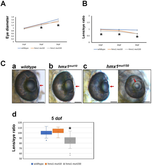
(A) Tracking of the growth of wildtype, hmx1mut10 and hmx1mut150 eye from 1 dpf to 3 dpf. Hmx1mut10 and hmx1mut150 eyes are reduced in size compared to wildtype embryos at 2 and 3 dpf stages. (B) Lens growth expressed as ratio lens/whole eye. hmx1mut10 and hmx1mut150 lenses are smaller at 3 dpf. (C) Wildtype, hmx1mut10 and hmx1mut150 eyes at 5 dpf. Wildtype and hmx1mut10 present a fully developed lens (5C, a-b), while hmx1mut150 embryos exhibit two opposing phenotypes (5C, c); on the left side a recovered lens is presented, while on the right a smaller lens presenting a clear phenotype called microphakia. (5C, d) graphic representation of lens/eye ratio; hmx1mut150 presents a wide range of distribution of the lens size. Red arrows indicate the lens at 5 dpf. Data are expressed as mean of three to 5 experiments. *; P<0.05 (ANOVA test). Bar, C 100 μm.

Expression Data

Expression Detail
Antibody Labeling
Phenotype Data
Fish:
Observed In:
Stage Range: Prim-5 to Day 5

Phenotype Detail
Acknowledgments
This image is the copyrighted work of the attributed author or publisher, and ZFIN has permission only to display this image to its users. Additional permissions should be obtained from the applicable author or publisher of the image. Full text @ PLoS One