|
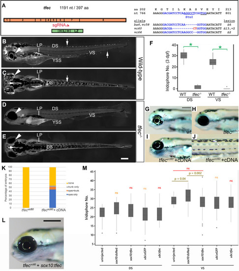
Loss of <italic>tfec</italic> function eliminates embryonic iridophores.(A) Schematic showing the distribution of the 8 exons of tfec (orange) in relation to the functional basic helix-loop-helix-leucine zipper domains (green) of the transcription factor. The red arrow indicates the position along both the gene and protein sequences targeted for mutagenesis by the CRISPR/Cas9 system. Included are the targeted WT tfec DNA/amino acid sequence (blue, with PAM underlined), and the sequences of the examined mutant alleles, with the corresponding molecular lesions (dashes for deleted nucleotides, red font for insertions). Imaging live embryos under reflected light reveals a striking lack of iridophores in tfec mutants (D,E) compared to their WT siblings (B,C) along the dorsal (downward arrow), ventral (upward arrow), and yolk sac stripes, as well as overlying the eye (arrowhead). Iridophores are also absent on the dorsal head (C,E, horizontal arrow) and the lateral patches. (F) Quantitation of differentiated iridophores across the dorsal and ventral trunk at 3 dpf confirms a prominent lack of iridophores along the dorsal and ventral stripe of tfec mutants. (G-K) Injection of tfec cDNA can rescue the mutant phenotype. Differentiated iridophores (arrowheads) are abundant on the eye of WT embryos (G), but completely absent from the eye of a tfecvc60 mutant sibling (H) at 4 dpf. Co-injection with Tol2 transposase of a construct where the tfec promoter drives transcription of the tfec cDNA sequence, leads to rescue of iridophores (arrowheads) on the eye (I) and trunk (J) of tfecvc60 mutants. (K) Quantitation of rescue efficiency. Approximately 45% of mutants displayed eye iridophore rescue, 3% showed rescue in the trunk only and 6% showed rescue both in the eyes and trunk (n = 62). By contrast, iridophores were never observed in uninjected tfecvc60 sibling larvae (n = 58). (L) tfec cDNA expressed from the sox10 promoter is capable of rescuing iridophores (arrowheads) in tfecvc60 mutants by 4 dpf. (M) At 3 dpf, numbers of iridophores in the dorsal stripe (DS) of wild-type embryos does not significantly change between uninjected embryos and embryos injected with either of the control constructs (sox10:dsRed, ubi:eGFP; orange “ns”). Embryos injected with sox10:tfec or with ubi:tfec show no significant change in DS iridophore numbers compared to both uninjected, and control injected siblings (sox10:dsRed, ubi:eGFP, respectively; red “ns”). In the ventral stripe (VS), injection of the ubi:tfec construct led to no significant iridophore number alterations, when compared to both control-injected and uninjected siblings (red “ns”). ubi:eGFP-injected controls do not show differences in VS iridophores when compared to uninjected controls (orange “ns”). When injecting sox10:dsRed, a weakly significant increase in VS iridophores is observed (p = 0.04). sox10:tfec injection does not lead to significant changes compared to uninjected controls, but appears to lead to a decrease in numbers when compared to sox10:dsRed (p = 0.002). Dots indicate outliers. sgRNA, small guide RNA; DS, dorsal stripe; VS, ventral stripe; LP, lateral patches; YSS, yolk sac stripe. (B,D, G-J,L): lateral views. (C,E): dorsal views. Head towards the left. Scale bars: 200 μm. (F): spots signify outlier values *, p-value < 10−9 using t-test.
|