
Figure 4
- ID
- ZDB-FIG-210128-161
- Publication
- Ozalp et al., 2021 - Nradd Acts as a Negative Feedback Regulator of Wnt/β-Catenin Signaling and Promotes Apoptosis
- Other Figures
- All Figure Page
- Back to All Figure Page
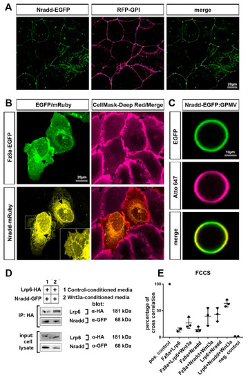
Nradd localizes to the plasma membrane and interacts with the Wnt–receptor complex (A) Nradd-EGFP (green) and RFP-GPI (red) localize at the plasma membrane of the enveloping layer cells of zebrafish embryos at 4 hpf (dome stage). (B) In U2OS cells, Fz8a-EGFP (green) co-localizes with the CellMask Deep Red (red) at the plasma membrane while Nradd-mRuby (yellow) localizes to the ER, but a significant fraction of it is in the plasma membrane, as seen from the zoom-in inset image. Arrows indicate protein transport from the ER to the plasma membrane. (C) Nradd-EGFP (green) localizes to the surface of cell-derived giant plasma membrane vesicles (GPMVs) and overlaps with the plasma membrane marker Atto647N-PE (red). (D) Nradd-GFP co-immunoprecipitates with Lrp6-HA in HEK293T cells. Three independent experiments were performed. (E) Fluorescence cross-correlation spectroscopy (FCCS) measurements show cross-correlation percentages between Fz8a-Lrp6, Fz8a-Nradd and Lrp6-Nradd, with/without Wnt3a stimulation at the plasma membrane. Nradd co-diffuses with Fz8a and Lrp6 at the membrane and this co-diffusion is enhanced by Wnt3 stimulation. EGFP and mCherry proteins were used as negative controls and their cross-correlation percentage is 1.70%. |