
Figure 2.
- ID
- ZDB-FIG-210119-22
- Publication
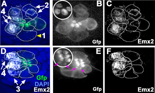
- Ohta et al., 2020 - Emx2 regulates hair cell rearrangement but not positional identity within neuromasts
- Other Figures
- All Figure Page
- Back to All Figure Page
( |
| Gene: | |
|---|---|
| Antibody: | |
| Fish: | |
| Anatomical Terms: | |
| Stage: | Day 4 |