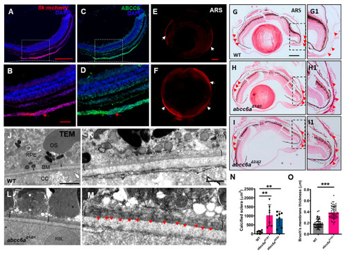
abcc6a mutants display ectopic calcification in the eyes. (A–D) Immunofluorescence results show that Abcc6 is located in the vascular-rich choroidal tissues by co-localization staining (red arrow) of Tg (flk: mcherry) fish. (E,F) Whole mount Alizarin Red staining shows ectopic scleral calcification in isolated eyes of abcc6aΔ1/Δ1 mutants. (G–I; G1–I1) Alizarin Red staining shows that the scleral layer of the abcc6aΔ1/Δ1 and abcc6aΔ2/Δ2 mutant eyes displays an accumulation of abnormal calcification (H,I; H1,I1) compared to WT (G,G1). (G1–I1) Higher-magnification images of the dashed boxes in (G–I). Red arrow marks abnormal calcification of scleral layer in the abcc6aΔ1/Δ1 mutant eyes. (J–M) Transmission electron microscopy showing abnormal thickening and enrichment of dense electron core of Bruch’s membrane in abcc6aΔ1/Δ1 mutant eyes (L), compared with WT eyes (J). (K,M) Higher-magnification images of the dashed boxes in (J,L). Black brackets indicate the position of BM, and red arrow indicates position of dense electron core. RPE, retinal pigment epithelium; BM, Bruch’s membrane; CC, choriocapillaris; OS, outer segment; RBC, red blood cell. (N) Quantification of area of calcification of sclera of WT (n = 7), abcc6aΔ1/Δ1 (n = 7) and abcc6aΔ2/Δ2 fish (n = 7). ** p < 0.001, Student’s t-test (unpaired, two-tailed). (O) Quantification of Bruch’s membrane thickness of WT (n = 6, 10 measuring points per sample) and abcc6aΔ1/Δ1 (n = 6, 10 measuring points per sample) fish. *** p < 0.001, Student’s t-test (unpair, two-tailed). Scale bar: 250 μm (A,C; E,F; G–I); 200 μm (B,D);62.5 μm (G1–I1); 2 μm (J,L); 0.5 μm (K,M).
|
