FIGURE

Fig 4

ID
ZDB-FIG-210113-119
Publication
Nunley et al., 2020 - Defect patterns on the curved surface of fish retinae suggest a mechanism of cone mosaic formation
Other Figures
All Figure Page
Back to All Figure Page
Fig 4

By photoconverting UV cones near the retinal margin, we track Y-Junction motion.

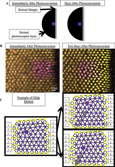
(A) Schematic of photoconverted UV cones in photoreceptor epithelium near the retinal margin. We photoconvert a patch of UV cones (purple box) near the margin, where new cone photoreceptors are incorporated by mitotic addition. After two, three or four days of retinal growth, we image the photo-converted region, which is now separated from the margin by newly added retinal tissue (black). (B) Example of patch of UV cones immediately after photoconversion and two days later. In this line (Tg[sws1:nEOS]), UV cones express a nuclear-localized, photoconvertible fluorescent protein under control of the UV cone opsin promoter. The non-photoconverted fluorescent protein is pseudo-colored yellow, and the photoconverted fluorescent protein is pseudo-colored magenta. Retinal margin is to the right of each image. Approximately eight columns of UV cones have been added in two days since photoconversion. (C) Glide motion involves subtle motion of individual UV cones near the Y-junction core. Magenta circles are UV cones with photoconverted fluorescent signal, and yellow circles are surrounding UV cones with non-photoconverted fluorescent signal. Every cone is connected to nearest neighbors. Five-sided and seven-sided stars: five-coordinated and seven-coordinated UV cones, respectively. Dashed black line: the “inserted” row. The two triangulations on the right describe positions of UV cones (from the left triangulation) after glide in the direction denoted by gray arrow. Note that the assignment of five- and seven-coordinated UV cones has shifted by one row.

Expression Data

Expression Detail
Antibody Labeling
Phenotype Data

Phenotype Detail
Acknowledgments
This image is the copyrighted work of the attributed author or publisher, and ZFIN has permission only to display this image to its users. Additional permissions should be obtained from the applicable author or publisher of the image. Full text @ PLoS Comput. Biol.