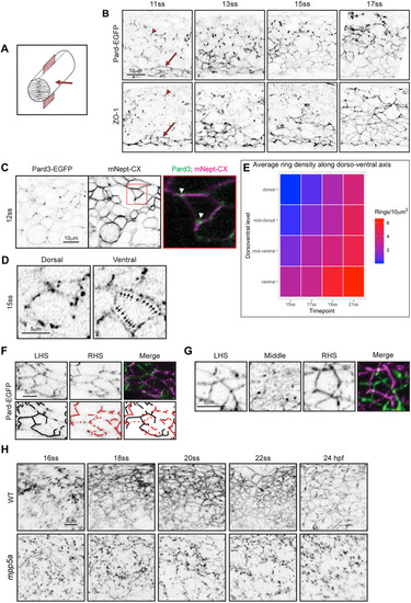
Mpp5a-dependent transition from spot adhesion to apical ring. (A) Diagram of neural rod with inserted red sheet to illustrate sagittal plane of confocal sections. Red arrow indicates direction of imaging. (B) Sagittal confocal planes of neural rod in anterior spinal cord region at 11-, 13-, 15- and 17-somite stages (ss). Comparable images were seen at each time point from three embryos. Images in the bottom row are from fixed embryos stained for ZO-1. Comparable images were seen from two embryos at each time point. Red arrow indicates ventral apical rings and red arrowhead indicates puncta. Dorsal is at the top of each panel. (C) Sagittal confocal images at 12-somite stage showing that Pard3-EGFP puncta are often located at cell vertices (arrowheads, n=8 embryos). Plasma membrane was imaged using mNeptune2.5-CAAX (mNept-CX). (D) Examples of punctate Pard3-EGFP in developing apical rings from a 15-somite-stage embryo. Dorsal example shows an immature, incomplete ring. Ventral example shows a more complete ring, with arrows indicating multiple puncta between vertices. Single sagittal confocal planes. (E) A heat-map quantification of the formation of mature Pard3 apical rings from two embryos over developmental time and dorsoventral position. (F) Parasagittal confocal sections from left-hand side (LHS) and right-hand side (RHS) of 15-somite stage neural rod. Incomplete apical rings of Pard3-EGFP are forming independently on left and right sides of the midline. (G) Parasagittal and sagittal confocal sections at 17-somite stage showing complete apical rings of Pard3-EGFP on either side of the midline. The sagittal section (labelled Middle) shows a largely diffuse, low level of Pard3-EGFP expression in the midline territory between the left and right rings (see Movie 1). Apical ring formation in F,G was analysed from >10 embryos. For each embryo, 10-25 apical rings were located on one side of the neuroepithelium, and a z-stack was taken through the middle of the neural rod until the rings on the opposite side were visible. (H) Images taken from confocal time-lapse movies of wild-type (WT) and mpp5a morphant Pard3-EGFP embryos in sagittal orientation from the 16-somite stage to 24 hpf. Comparable images were obtained from three embryos from each genotype. Three control morphants were also assessed in the Cdh2-tFT transgenic line, and all had apical rings comparable to wild types.
|
