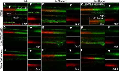
Cya and BMS-833923 both block Hh signaling in zebrafish larvae. A–I, Lateral views of the trunk of TgBAC(ptch2:Kaede) larvae showing Kaede expression in Hh-responsive cells of the ventral spinal cord at different time points after UV photoconversion. Smaller panels show separated color channels. A, D, G, The border of native (green) and photoconverted (red) Kaede protein in the spinal cord immediately after photoconversion of the anterior trunk region. B, C, In DMSO-treated control larvae newly synthesized Kaede protein (arrowheads) was easily detectable after 24 h and continued to increase through 48 and 72 h, when converted and non-converted protein levels were very roughly equivalent (C, inset). E, F, Treatment with 5 μm BMS-833923 effectively blocked the synthesis of new Kaede protein (arrowheads) in the ventral spinal cord at 24 h (E), with low levels of newly synthesized Kaede protein becoming visible after 48 h at this drug concentration (F). H, I, Treatment with 10 μm Cya effectively blocked new Kaede synthesis (arrowheads) for 24 h (H), with low levels of expression being visible after 48 h at this low Cya concentration (I).
|
