FIGURE

Figure 1

ID
ZDB-FIG-201230-38
Publication
Epting et al., 2020 - Loss of CBY1 results in a ciliopathy characterized by features of Joubert syndrome
Other Figures
All Figure Page
Back to All Figure Page
Figure 1

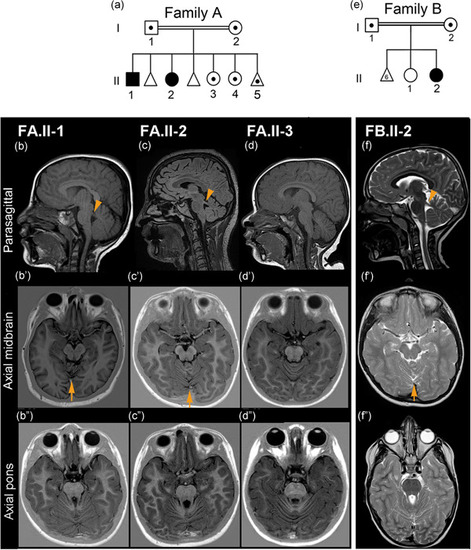
Pedigree of Families A and B and cerebral MRI findings. (a) Pedigree of Family A showing the two affected siblings (black symbols) and carriers (symbols with the dot) of the CBY1 variant Chr22:g.39067079_39067080del within the family. The first two fetuses were not genotyped. (b−f) Cerebral magnetic resonance imaging examinations in Family A (FA.II‐1, FA.II‐2, FA.II‐3), and Family B (FB.II‐2). In the parasagittal views (b,c,f), the superior cerebellar peduncles (arrowheads) are more horizontally oriented, as opposed to the normal and more vertically oriented peduncle in (d). In the axial midbrain views (bʹ,cʹ,fʹ), cerebellar vermian foliar dysplasia is seen above the arrows. This anomaly is not present in (dʹ). The axial pons views (b″,c″,f″) show elongation of the superior cerebellar peduncles giving a mild “molar tooth” appearance, but not in (d″). Sagittal views (b–d) are T1 and (f) T2 weighted. Axial midbrain and pons views (bʹ–dʹ) and (b″–d″) are inversion recovery and (f–f″) are T2 weighted. (e) Pedigree of Family B showing the affected sibling and her parents, which are carriers of the CBY1 variant Chr22:g.39064123_39064124dup. FB.II‐1 and the fetuses were not genotyped

Expression Data

Expression Detail
Antibody Labeling
Phenotype Data

Phenotype Detail
Acknowledgments
This image is the copyrighted work of the attributed author or publisher, and ZFIN has permission only to display this image to its users. Additional permissions should be obtained from the applicable author or publisher of the image. Full text @ Hum. Mutat.