FIGURE

Fig. 5

ID
ZDB-FIG-201212-17
Publication
Issaka Salia et al., 2020 - Bioinformatic analysis and functional predictions of selected regeneration-associated transcripts expressed by zebrafish microglia
Other Figures
All Figure Page
Back to All Figure Page
Fig. 5

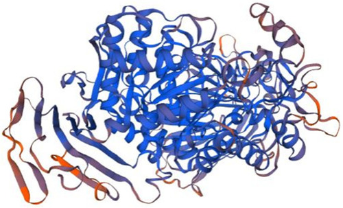
Homology model of P11. Maltase-glucoamylase, intestinal (3top.1.A in the rcsb protein database) is the template used for the homology modelling of P11. The X-RAY diffraction 2.9 Å was used to determine the experimental structure of 3top.1.A [63]. The homology model starts from P11 residue N°922 (LYS, Lysine) and ends with the residue N° 1804 (PHE, Phenylalanine). The P-type trefoil domain (amino acid N°51–962), galactose mutaros domain (amino acid N°114–1085), and glycoside hydrolase domain (amino acid N°225–1152) are not covered in the homology model. The blue color show regions of the model where P11 was well modeled and orange regions show where P11 was poorly modeled

Expression Data

Expression Detail
Antibody Labeling
Phenotype Data

Phenotype Detail
Acknowledgments
This image is the copyrighted work of the attributed author or publisher, and ZFIN has permission only to display this image to its users. Additional permissions should be obtained from the applicable author or publisher of the image. Full text @ BMC Genomics