FIGURE

Figure 3

ID
ZDB-FIG-201102-21
Publication
Santhanam et al., 2020 - A Zebrafish Model of Retinitis Pigmentosa Shows Continuous Degeneration and Regeneration of Rod Photoreceptors
Other Figures
All Figure Page
Back to All Figure Page
Figure 3

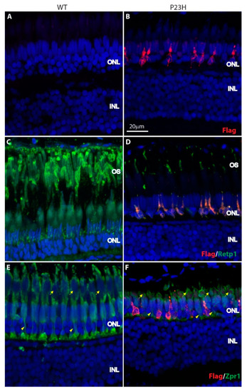
Expression of P23H Flag-tagged rhodopsin in adult zebrafish retina. (A,B) The Flag-tagged P23H mutant rhodopsin (red) is expressed by sparsely-distributed photoreceptors in the adult fish (B); there is no labeling in wild type fish (A). (C,D) Mutant rhodopsin (red) colocalizes with rhodopsin (Retp1, green) in the rods. (E,F) Mutant rhodopsin (red) is expressed only by the rods and not the cones, as seen with the cone marker Zpr1 (green). Yellow arrowheads represent the cone axons and yellow arrows represent the cone myoids. ONL: outer nuclear layer; INL: inner nuclear layer; OS: outer segments. Scale bar in B applies to all panels.

Expression Data

Expression Detail
Antibody Labeling
Phenotype Data
Fish:
Observed In:
Stage: Adult

Phenotype Detail
Acknowledgments
This image is the copyrighted work of the attributed author or publisher, and ZFIN has permission only to display this image to its users. Additional permissions should be obtained from the applicable author or publisher of the image. Full text @ Cells