
FIGURE
Fig. 4
- ID
- ZDB-FIG-201029-8
- Publication
- Bagnat et al., 2020 - Development of a straight vertebrate body axis
- Other Figures
- All Figure Page
- Back to All Figure Page
Fig. 4
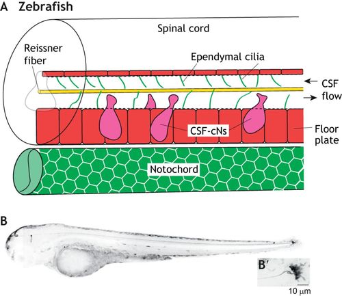
The integration of the Reissner fiber and CSF-resident components. (A) Schematic of the larval zebrafish spinal cord and notochord, showing the close associations of the Reissner fiber (yellow) with ependymal motile cilia (green lines) and CSF-cNs (magenta). The floor plate is shown in red. (B) Inverted grayscale confocal image of a 3 days post-fertilization larval scospondin-GFPut24 knock-in line in a lateral view. (B’) High magnification image of the terminal ampulla region. Taken from Troutwine et al., 2019 preprint. |
Expression Data
Expression Detail
Antibody Labeling
Phenotype Data
Phenotype Detail
Acknowledgments
This image is the copyrighted work of the attributed author or publisher, and
ZFIN has permission only to display this image to its users.
Additional permissions should be obtained from the applicable author or publisher of the image.
Full text @ Development