FIGURE

Fig. 6

ID
ZDB-FIG-201003-239
Publication
Corallo et al., 2020 - Autophagic flux inhibition enhances cytotoxicity of the receptor tyrosine kinase inhibitor ponatinib
Other Figures
All Figure Page
Back to All Figure Page
Fig. 6

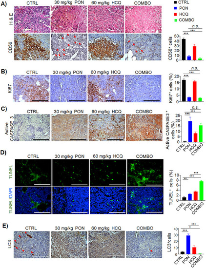
Histopathological examination of post-therapy neuroblastoma tumors confirms the efficacy of combination treatment in vivo. a Hematoxylin & Eosin (H&E) of tumor tissues and immunohistochemistry staining of CD56 proteins were carried out. Red arrows indicate positive cellular labeling. Scale bar, 150 μm. b Immunohistochemistry staining of Ki67, c active CASPASE 3, and d TUNEL assay for the detection of proliferating and apoptotic cells, respectively. The percentage (%) of positive events is reported in the histograms on the right. Scale bar, 150 μm. e Immunohistochemistry staining of LC3-positive cells in resected post-therapy tumors. Red arrows indicate positive cellular labeling. Scale bar, 150 μm. The percentage (%) of LC3-positive cells is reported in the histogram on the right. *p < 0.05, **p < 0.01, ***p < 0.001

Expression Data

Expression Detail
Antibody Labeling
Phenotype Data

Phenotype Detail
Acknowledgments
This image is the copyrighted work of the attributed author or publisher, and ZFIN has permission only to display this image to its users. Additional permissions should be obtained from the applicable author or publisher of the image. Full text @ J. Exp. Clin. Cancer Res.