FIGURE

Figure 8

ID
ZDB-FIG-201003-146
Publication
Head et al., 2020 - Vitamin E is necessary for zebrafish nervous system development
Other Figures
All Figure Page
Back to All Figure Page
Figure 8

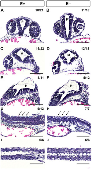
Histological analysis of 24 hpf zebrafish embryos with morphological defects associated with VitE status. Hematoxylin and eosin staining of E+ and E− embryos at 24 hpf was used to evaluate morphological defects, including fore- (F) and mid- (M), and hind- (H) brain ventricle inflation, somite formation and notochord vacuolation. Transverse section of E+ embryos had tear-drop shaped-F with eyes to each side (A), whereas E− embryos had a neural lumen, but improperly inflated-F (B). Serial transverse sections of E+ embryos showed normal M ventricle inflation with distinct hinge-points, indicated by *(C). E− embryos (D) had reduced M ventricle inflation with similar hinge points. E+ and E− embryos had similar H inflation leading to the neural tube in the trunk (E, F). Sagittal sections of the trunk indicate defined somite epithelial boundaries in E+ embryos (G) and indistinct epithelial boundaries in E− somitic cells (H). Notochord vacuolation appeared normal in both E+ (I) and E− (J) embryos. Scale bar represents 50 μm; representative embryos are shown. Figure panels were generated with the BZ- × 700 microscope, processed with BZ-X Analyzer Software with image adjustments equally applied across time points in Adobe Photoshop, v21.2.1.

Expression Data

Expression Detail
Antibody Labeling
Phenotype Data
Fish:
Condition:
Observed In:
Stage: Prim-5

Phenotype Detail
Acknowledgments
This image is the copyrighted work of the attributed author or publisher, and ZFIN has permission only to display this image to its users. Additional permissions should be obtained from the applicable author or publisher of the image. Full text @ Sci. Rep.