
Fig. 4
- ID
- ZDB-FIG-200924-44
- Publication
- Rodríguez-Morales et al., 2020 - Expression patterns of activating transcription factor 5 (atf5a and atf5b) in zebrafish
- Other Figures
- All Figure Page
- Back to All Figure Page
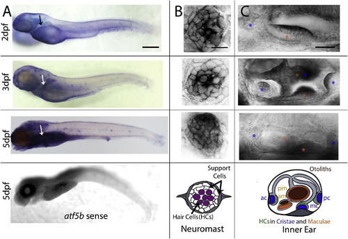
Larval expression of atf5b. (A) Whole larva at 2, 3 and 5dpf hybridized with an antisense RNA probe against the atf5b gene (3 top panels) and a sense probe (last panel). Black and white arrows indicate the developing liver. (B) High magnification of neuromasts (NMs) in 2, 3 and 5dpf larvae (top panels), which are schematized (bottom panel) with centrally located hair cells HCs (magenta) surrounded by two type of support cells (white and grey). (C) lateral views of the inner ear (schematized in bottom panel) of a 2, 3 and 5dpf larva (top panels), showing strong atf5b expression in the sensory patches of the inner ear, namely in the anterior (ac), median (mc) and posterior (pc) cristae (blue asterisks) as well as the anterior (am) and posterior (pm) maculae (orange asterisks). Scale bars: in A = 200 μm, in B = 40 μm, and in C = 75 μm. |
Reprinted from Gene expression patterns : GEP, 37, Rodríguez-Morales, R., Vélez-Negrón, V., Torrado-Tapias, A., Varshney, G., Behra, M., Expression patterns of activating transcription factor 5 (atf5a and atf5b) in zebrafish, 119126, Copyright (2020) with permission from Elsevier. Full text @ Gene Expr. Patterns