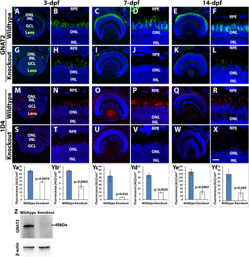
Cone outer segment generation is reduced in tmem216 knockout fish. Cryosections of whole zebrafish heads were stained with cone OS markers GNAT2 (green) and 1D4 (red) at 3-, 7- and 14-dpf. All sections were counterstained with DAPI to visualize nuclei. Knockout panels shown were from the tmem216snyR8Δ60 line. Similar results were observed in the tmem216snyΔ175 line. (A–F) GNAT2 immunostaining of wild-type retina at 3-, 7-, and 14-dpf, respectively. (G–L) GNAT2 staining of tmem216snyR8Δ60 homozygous retina at 3-, 7-, and 14-dpf, respectively. (M–R) 1D4 immunostaining of wild-type retina at 3-, 7-, and 14-dpf, respectively. (S–X) 1D4 labeling of tmem216snyR8Δ60 homozygous retina at 3-, 7-, and 14-dpf, respectively. (Ya, Yc, Ye) Quantification of GNAT2 fluorescence intensity between wildtype and knockout retinas at 3-, 7-, and 14-dpf, respectively. (Yb, Yd, Yf) Quantification of 1D4 fluorescence intensity between wildtype and tmem216snyR8Δ60 homozygous retina at 3-, 7- and 14-dpf, respectively. Note that GNAT2 and 1D4 immunoreactivity was significantly reduced at 3-, 7- and 14-dpf (n = 3, Student's t-test). (Z) Western blotting using anti-GNAT2 was performed on whole head lysates collected from three 7-dpf wildtype and tmem216snyR8Δ60 homozygous zebrafish larvae. The intensity ratio of GNAT2/-β-actin between wildtype and knockout was reduced from 0.88 to 0.27. Scale bar in X: 55 µm for A, G, M, and S; 110 µm for C, I, E, K, O, U, Q, and W; 6 µm for B, H, D, J, F, L, N, T, P, V, R, and X. GCL, ganglion cell layer; INL, inner nuclear layer; ONL, outer nuclear layer; RPE, retinal pigment epithelium.
|
