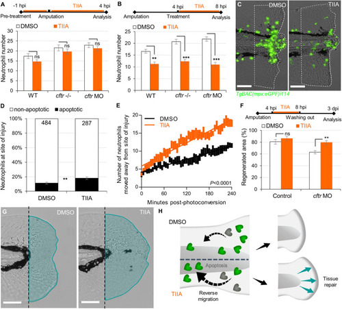
TIIA-driven neutrophil apoptosis and reverse migration accelerate inflammation resolution in CF. (A)TgBAC(mpx:EGFP)i114 larvae were pretreated with 25 μM of TIIA prior to tail fin amputation procedure, then injured and immediately put back in treatments for 4 h. Neutrophil number at the wound was counted at 8 hpi (n = 21, two-tailed Bonferroni t-test). (B,C)TgBAC(mpx:EGFP)i114 larvae were injured and treated from 4 hpi with 25 μM of TIIA. (B) Neutrophil number at wound was counted at 8 hpi (n = 21, two-tailed Bonferroni t-test). (C) representative number of neutrophils remaining at wounds at 8 hpi (scale bars, 200 μm). (D) Neutrophil apoptosis quantification at 8 hpi in cftr MO treated with 25 μM of TIIA from 4 hpi and stained with TUNEL/TSA. (n = 40, Fisher t-test). (E) Reverse-migration assay in cftr MO Tg(mpx:gal4)sh267;Tg(UASkaede)i222. At 4 hpi fish were treated with 25 μM of TIIA and neutrophils at site of injury were photoconverted. The numbers of photoconverted cells that moved away from the wound were time-lapse imaged and quantified over 4 h. (F,G) Regenerative performance after TIIA treatment. (F) Regenerated fin areas are measured at 3 dpi (n = 21, two-tailed Bonferroni t-test). (G) Representative imaging of injured tail fin at 3 dpi (scale bars, 200 μm). (H) Schematic diagram showing TIIA efficiently accelerates inflammation resolution by inducing neutrophil apoptosis and reverse migration at wounds and improves tissue repair in CF animal.
|
