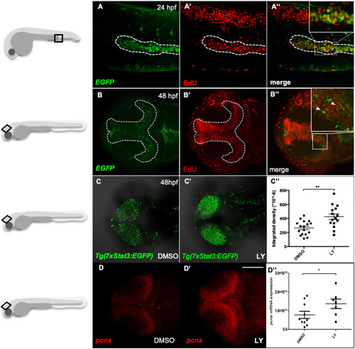
Stat3 pathway is active in proliferating cells of zebrafish haematopoietic tissue and optic tectum. (A-A″) Fluorescence co-localization (A″) using α-EGFP Ab (A) and EdU proliferation assay (A′) in the haematopoietic tissue (dashed line) of 22 hpf Tg(7xStat3:EGFP) reporter embryos. (B-B″) Fluorescence co-localization (B″) between fish using α-EGFP probe (B) and EdU proliferation assay (B′) in the optic tectum (TeO; dashed line) of 48 hpf embryos. (C,C′) In vivo fluorescence of Tg(7xStat3:EGFP) reporter activity in the TeO of embryos treated with LY364947 inhibitor between 24 and 48 hpf (C′) compared with DMSO-treated controls (C). (C″) Relative fluorescence intensity in the TeO of 48 hpf Tg(7xStat3:EGFP) embryos described in C (n=15, P=0.0014). (D,D′) Whole mount in situ hybridization detection of pcna mRNA in the TeO of 48 hpf embryos treated with LY364947 inhibitor between 24 and 48 hpf (D′) compared with DMSO-treated controls (D). (D″) pcna mRNA expression in the embryos described in D (P=0.038). All statistical analyses were performed by unpaired t-test; *P<0.05, **P<0.01. Graphs indicate mean±s.e.m. Scale bar: 100 μM.
|
