
Fig. 1
- ID
- ZDB-FIG-200709-55
- Publication
- Harris et al., 2020 - Long-Range Optogenetic Control of Axon Guidance Overcomes Developmental Boundaries and Defects
- Other Figures
- All Figure Page
- Back to All Figure Page
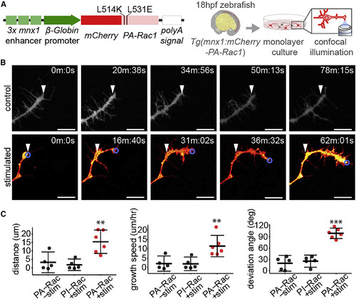
Optogenetic Stimulation of PA-Rac1 Directs Axonal Growth of Cultured Zebrafish Spinal Motor Neuron Axons (A) Schematic of expression construct and experimental design. (B) Time series of cultured PA-Rac1+ neurons, either unstimulated (top row) or illuminated in a region of interest focused on the leading edge of the growth cone (blue circle) (bottom row). White arrowheads indicate the initial position of the growth cone (scale bar, 10 μm). (C) Stimulated axons (n = 5 axons from separate cultures of independent embryos) grew significantly greater distances over the trial period (left), resulting in a faster rate of growth (middle), and deviated significantly from their initial trajectory compared with unilluminated PA-Rac1+ axons or illuminated photo-insensitive Rac1+ (PI-Rac1) axons (right, independent samples Student’s t test, ∗∗p < 0.01, ∗∗∗p < 0.001. Mean and 95% CIs shown). See also Figure S1; Video S1. |
Reprinted from Developmental Cell, 53, Harris, J.M., Wang, A.Y., Boulanger-Weill, J., Santoriello, C., Foianini, S., Lichtman, J.W., Zon, L.I., Arlotta, P., Long-Range Optogenetic Control of Axon Guidance Overcomes Developmental Boundaries and Defects, 577-588.e7, Copyright (2020) with permission from Elsevier. Full text @ Dev. Cell