
FIGURE
Fig. 6
- ID
- ZDB-FIG-200708-13
- Publication
- Wu et al., 2020 - Impaired oocyte maturation and ovulation in membrane progestin receptor (mPR) knockouts in zebrafish
- Other Figures
- All Figure Page
- Back to All Figure Page
Fig. 6
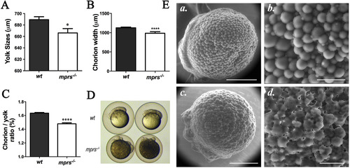
Malformed embryos from in all seven membrane progestin receptor paralogues mutant (mprs−/−) females. (A–C) Reduced yolk size, chorion width, and the ratio of chorion size to yolk size in the mprs−/−embryos. 30% epiboly embryos were used (n = 7). (D) Representative malformed embryos (16-cell stage) from mprs−/− females with smaller yolk and chorion. (E) Disorganized and scrambled yolk granules on the surface of mprs−/− early stage embryo (32-cell stage embryo). a & b) wt; c & d) mprs−/−. Scale bars: a & c) 200 μm; b & d) 50 μm. |
Expression Data
Expression Detail
Antibody Labeling
Phenotype Data
| Fish: | |
|---|---|
| Observed In: | |
| Stage Range: | 1-cell to 30%-epiboly |
Phenotype Detail
Acknowledgments
This image is the copyrighted work of the attributed author or publisher, and
ZFIN has permission only to display this image to its users.
Additional permissions should be obtained from the applicable author or publisher of the image.
Reprinted from Molecular and Cellular Endocrinology, 511, Wu, X.J., Liu, D.T., Chen, S., Hong, W., Zhu, Y., Impaired oocyte maturation and ovulation in membrane progestin receptor (mPR) knockouts in zebrafish, 110856, Copyright (2020) with permission from Elsevier. Full text @ Mol. Cell. Endocrinol.