|
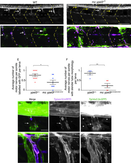
Ypel3 is required for perineurial glia ensheathing.(A) WT larva at 56 hpf. Perineurial glia (expressing nkx2.2a:GFP, green) have migrated out of the CNS and begun to ensheath the motor nerve. White arrows indicate perineurial glia associated with the immature motor nerve. (B) mz ypel3-/- mutant at 56 hpf. At this stage, perineurial glia are largely absent, and the few that migrate out of the CNS appear wispy and thin (yellow arrows). (C) In WT larvae at 5 dpf, perineurial glia are tightly associated with Schwann cells (expressing sox10:mRFP, magenta) forming slender tubes (white arrows). A small percentage of WT perineurial cells have not formed these slender tubes (arrowhead). (D) In mz ypel3-/- mutants, at 5 dpf, perineurial glia are overgrown and fail to associate with the Schwann cells. Yellow dashed lines label the somite boundaries. Scale bar: 25 μm. (E-F) Quantification of the average number of motor nerves per somite labeled with GFP (E) and the average number of motor nerves per somite with slender tube morphology (F). (G-G”) WT larvae at 7 dpf. Perineurial glia have completely wrapped the Schwann cells forming a tubular structure (arrows). Some mRFP expression remains within the Schwann cells. (G) Merge. (G’) mRFP expression is clustered within the GFP positive domain (outlined with white dashed lines), which is composed of perineurial cells. (G”) Perineurial cells form a tubular structure (outlined with white dashed lines). (H-H”) mz ypel3 mutant at 7 dpf. Perineurial glia are overgrown and form a defasciculated structure. (H) Merge. (H’) mRFP signal is elevated in the mz ypel3 mutant Schwann cells and delineates three defasciculated structures (1, 2, 3). (H”) mz ypel3 mutant perineurial glia fail to wrap Schwann cells correctly resulting in an enlarged nerve (outlined with white dashed lines). White horizontal line in (G-H”) spinal cord ventral border. White horizontal dashed line (A-D) horizontal myoseptum. SC: spinal cord. *: loose perineurial cells. All images are lateral views. Scale bars: 25 μm in A-D, 5 μm in G-H”.
|