
FIGURE
Fig. 4
Fig. 4
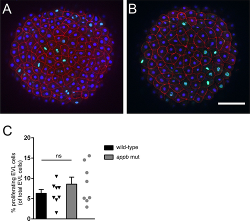
Proliferation of EVL in wild-type and appb mutants at 4 hpf. Representative confocal images of the EVL in wild-type (A) and appb mutant (B) embryos immunostained for the mitotic marker phospho-Histone H3 (pHH3; green) and actin binding phalloidin (red) at 4 hpf. All nuclei were labeled with DAPI (blue). Number of pHH3-positive EVL nuclei shown in percentage of all EVL cells (C). Student’s t-test was performed on wild-type (n = 8) and appbmutant (n = 8). Scale bar, 100 μm. |
Expression Data
Expression Detail
Antibody Labeling
Phenotype Data
| Fish: | |
|---|---|
| Observed In: | |
| Stage: | Sphere |
Phenotype Detail
Acknowledgments
This image is the copyrighted work of the attributed author or publisher, and
ZFIN has permission only to display this image to its users.
Additional permissions should be obtained from the applicable author or publisher of the image.
Full text @ Sci. Rep.