|
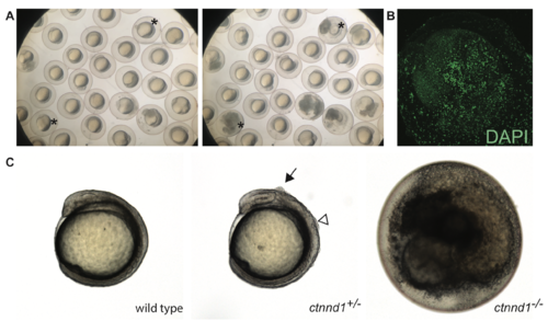
Embryonic disintegration in ctnnd1 mutants. (A) Time-lapse imaging illustrates disintegrating phenotype of ctnnd1 mutants. Asterisk(s) mark two of the embryos that disintegrated during the time-lapse recording. (B) DAPI staining of fixed ctnnd1 mutant embryos reveals that cells with intact nuclei dissociate from the embryo. (C) Phenotypes of ctnnd1 sibling embryos at the ~6-somite stage. The homozygous mutant has disintegrated, the heterozygous mutant displays clumps of cells along the dorsal surface, and the wild-type embryo appears normal. The solid arrow marks a clump of cells dorsal to the midbrain and the open arrowhead marks a clump of cells dorsal to the hindbrain in the heterozygote.
|