|
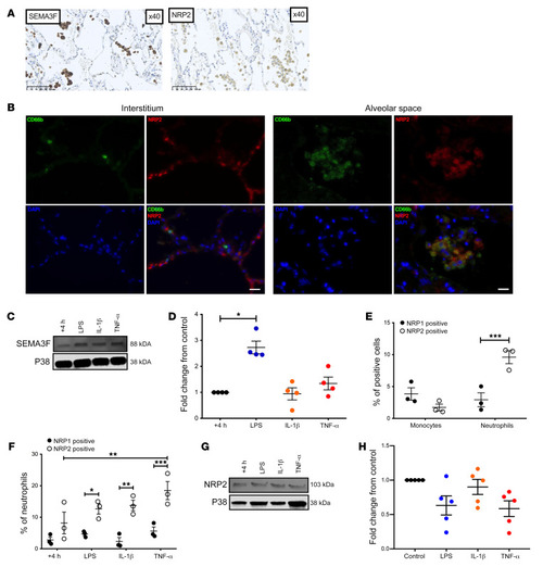
Inflammatory human neutrophils express SEMA3F and its coreceptor NRP2.(A and B) Lung sections taken at time of tumor resection from nontumor regions of patients with moderate severity COPD were stained for SEMA3F or NRP 2 (A), or a combination of CD66b (green), NRP2 (red), and DAPI (blue) (B). Images taken at ×40 magnification. Scale bars: 100 μm in A, 20 μm in B. Human blood neutrophil SEMA3F protein expression following 4 hours culture ex vivo was assessed by Western blot (C), and fold change to unstimulated control was determined by densitometry normalized to P38 (D). The percentage of blood monocytes (CD66b–, CD14/49D+) and neutrophils (CD66b+) expressing NRP1 and NRP2 was determined in freshly isolated cells (E) and following ex vivo culture for 4 hours by flow cytometry in control and stimulated conditions (F). Data are mean ± SEM, with individual data points (n = 3–5) from independent experiments. Human blood neutrophil NRP2 protein expression following 4 hours culture ex vivo was assessed by Western blot (G) and fold change to unstimulated control was determined by densitometry normalized to P38 (H). Statistical analysis: 1-way ANOVA and Bonferroni’s post hoc tests (D and H) and 2-way ANOVA and Sidak’s post hoc tests (E and F) were performed. *P < 0.05; **P < 0.01; ***P < 0.001.
|