
Fig. 1
- ID
- ZDB-FIG-200522-11
- Publication
- Bahrami et al., 2020 - Development of vascular regulation in the zebrafish embryo
- Other Figures
- All Figure Page
- Back to All Figure Page
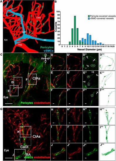
Mural cell morphology and coverage of the cerebral vasculature at 6 dpf. (A) A model of vSMC and pericyte locations on 6 dpf zebrafish embryo cerebral vessels. PHS, primordial hindbrain sinus; CaDI, caudal division of the internal carotid artery; BA, basilar artery; CtAs, central arteries. (B) Pericytes are typically found on vessels with diameters of ≤6.5 μm (n=215 pericyte-covered vessel regions), while vSMCs appear on the larger cerebral vessels (≥9.5 μm; n=130 vSMC-covered vessel regions). Cerebral vessels with diameters of 6.5-9.5 μm are covered by a mix of pericytes and vSMCs. (C) Lateral image of cerebral vessel pericyte coverage at 6 dpf. (D-F‴) Enlargements of vessels in C, highlighting contact between processes of different pericytes. Dotted lines outline pericyte cell bodies; arrows mark processes. (G) Lateral image of cerebral vessel vSMC coverage at 6 dpf, focusing on the CaDI and BA. (H-J‴) Enlargements of the vessels in G showing extensive vSMC coverage. Scale bars: 50 µm in C and G; 10 µm in D-F‴,H-J‴. A, P, V and D refer to anterior, posterior, ventral and dorsal. |