
Fig. 5
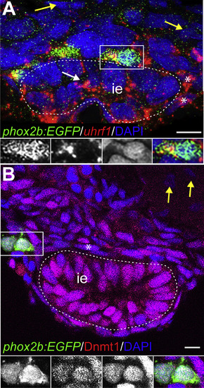
uhrf1 and Dnmt1 are expressed in enteric, epithelial, and smooth muscle progenitors during development. Transverse sections of stained embryos. (A) At 48 hpf, uhrf1 (red) is expressed in phox2b:EGFP (green) positive EPCs and also in DAPI-positive (blue) ISMPs (asterisks) and epithelial progenitors (white arrow). Yellow arrows point to uhrf1 negative cells outside of the nascent intestine. White dashed line indicates intestinal epithelium (ie). Insets show enlargement of cell (white box), GFP, uhrf1, DAPI, and overlay from left to right. Note that the GFP staining appears speckled due to the RNA in situ hybridization procedure. (B) At 48 hpf, Dnmt1 (red) is expressed in phox2b:EGFP (green) positive EPCs and in DAPI-positive (blue) ISMPs (asterisk). Insets show enlargements of outlined cell, GFP, Dnmt1, DAPI and overlay from left to right. Yellow arrow points to Dnmt1 negative cell outside of the intestine. Scale bar = 10 μm in A-B. |
| Genes: | |
|---|---|
| Fish: | |
| Anatomical Terms: | |
| Stage: | Long-pec |
Reprinted from Developmental Biology, 455, Ganz, J., Melancon, E., Wilson, C., Amores, A., Batzel, P., Strader, M., Braasch, I., Diba, P., Kuhlman, J.A., Postlethwait, J.H., Eisen, J.S., Epigenetic factors Dnmt1 and Uhrf1 coordinate intestinal development, 473-484, Copyright (2019) with permission from Elsevier. Full text @ Dev. Biol.