|
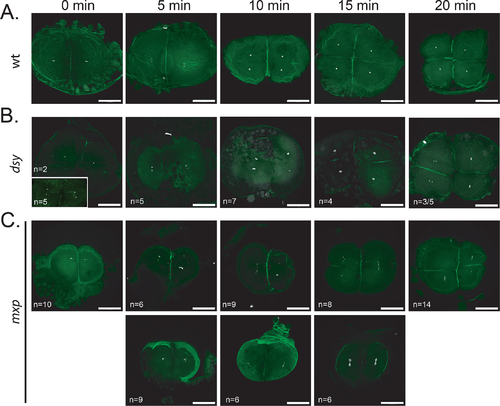
Examining nuclear integrity in mixed up and disarray embryos. A. Wild-type (TL), (B) dsy and (C) mxp embryos were fixed at 5-minute intervals spanning 20 minutes (corresponding to the 2 to 4 cell division) and stained with DAPI and phalloidin to mark the DNA and actin at the cell boundaries, respectively. B. Representative embryos (numbers indicated in the lower left corner) from a total of three dsy females. In some cases nuclear divisions were asynchronous (20 min) in embryos from dsy mutant mothers compared to wild type (A). C. Representative embryos (numbers indicated in the lower left corner) from a total of four mxp females. Embryos shown in the upper row underwent cell division timing similar to wild type in (A), whereas the embryos in the lower row were delayed. Scale bars = 200μm.
|