
Fig. 1
- ID
- ZDB-FIG-200505-9
- Publication
- Schwarzer et al., 2020 - Neurogenesis in the inner ear: the zebrafish statoacoustic ganglion provides new neurons from a Neurod/Nestin-positive progenitor pool well into adulthood
- Other Figures
- All Figure Page
- Back to All Figure Page
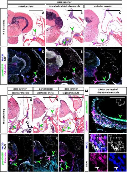
Anatomy of the statoacoustic ganglion (SAG) in the adult zebrafish. (A-L) H&E staining (A-C,G-I) and immunohistochemistry (D-F,J-L) of cross-sections through the entire inner ear show the innervation of the pars superior, the dorsal part of the inner ear consisting of the anterior (A,D), the lateral (B,E) and the posterior cristae (H,K) and the utricular macula (B,C,E,F) as well as the pars inferior, the ventral part of the inner ear including the saccular macula (G,J) and the lagenar macula (I,L) by the SAG, the eighth cranial nerve. Neurons of the SAG express the neuronal markers HuC/D (labelling neuronal cell bodies) and Calretinin [labelling neuronal cell bodies as well as the axons (purple arrowheads)]. Hair cells in the sensory patches are labelled using pou4f3:GFP (green arrowheads). The former position of the three otoliths lapillus (utricular macula), sagitta (saccular macula) and asteriscus (lagenar macula) are visible as light purple residual structures left after decalcification of the otoliths, close to the sensory hair cells in the H&E staining (A-C,G-I; marked with yellow asterisks). (M) Close-up of the medial part of the SAG at the level of the utricular macula; comparison of HuC/D with Calretinin reveals that the latter only labels a subpopulation of neurons in the dorso-lateral part of the SAG (green and white arrowheads point to HuC/D-positive but Calretinin-negative neurons). Bottom panels in M show magnification of boxed area in top panel. Scale bars: 200 µm for A-L; 50 µm for M. Cross-sections show dorsal to the top and lateral to the right. Distance from first section through semicircular canals to the section of interest: anterior cristae (A) 0.5 mm; lateral cristae (B) 1.39 mm; utricular macula (C) 1.66 mm; posterior cristae (G) 2.53 mm; saccular macula (H) 3.15 mm; lagenar macula (I) 3.44 mm. |