FIGURE

Fig 2

ID
ZDB-FIG-200412-9
Publication
Xie et al., 2020 - E2f5 is a versatile transcriptional activator required for spermatogenesis and multiciliated cell differentiation in zebrafish
Other Figures
All Figure Page
Back to All Figure Page
Fig 2

Spermatogenesis arrest in <italic>e2f5</italic> mutants.

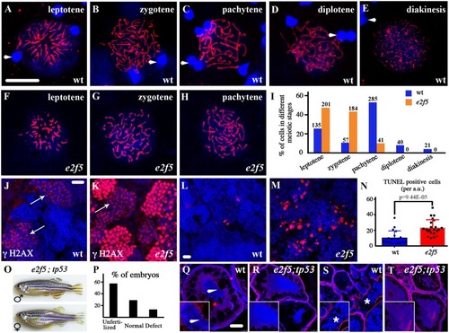
(A-H) Confocal images of primary spermatocytes at different prophase stages of meiosis I as indicated by anti-Sycp3 antibody staining (red). Arrowheads indicate spermatozoa nuclei from wild-type testis stained with DAPI in blue (A-E). (I) Bar graph showing the statistical results of the percentage of cells in different meiotic stages. The numbers of spermatocytes investigated are shown on top of each bar. (J-K) Staining of γH2AX (red) in the testes of wild-type and e2f5 mutant. Arrows point to the primary spermatocytes at leptotene and zygotene stages. (L-M) Confocal images showing apoptotic cells stained by TUNEL assay in red. (N) Bar graph with individual data points showing the number of TUNEL positive cells in wild-type and e2f5 mutant testes. (O) Phenotypes of e2f5;tp53 double mutants. (P) Bar graph showing the percentage of abnormal embryos produced from e2f5;tp53 double mutant females crossed with wild-type males. n = 441 from two double mutant females. (Q-T) Histological analysis of ovaries from wild-type and e2f5;tp53 double mutants. Inserted images are magnified views. Arrow in Q indicates peripheral localization of nucleoli stained by DAPI in stage II oocytes of wild-type ovary. The asterisk in S indicates cortical alveoli at later stage oocytes, which were visible under the confocal microscope due to autofluorescence. In panels J-M, Q-T, nuclei were counterstained with DAPI in blue. Scale bars: 10 μm in panel A-H, J-M and 50 μm in panel Q-T.

Expression Data

Expression Detail
Antibody Labeling
Phenotype Data
Fish:
Observed In:
Stage: Adult

Phenotype Detail
Acknowledgments
This image is the copyrighted work of the attributed author or publisher, and ZFIN has permission only to display this image to its users. Additional permissions should be obtained from the applicable author or publisher of the image. Full text @ PLoS Genet.