
Fig. 4
- ID
- ZDB-FIG-200331-15
- Publication
- Bhargava et al., 2019 - GCNA Preserves Genome Integrity and Fertility Across Species
- Other Figures
- All Figure Page
- Back to All Figure Page
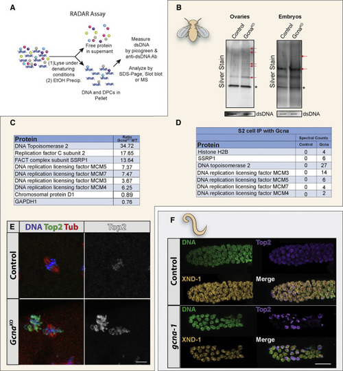
GCNA Mutants Exhibit Increased DPC Levels (A) Schematic illustrating principle of the RADAR assay. (B) RADAR was performed on Drosophila ovaries and embryos. Samples were normalized to dsDNA, separated by SDS-PAGE, and silver stained. Red arrows mark examples of differentially enhanced bands in the Gcna mutant samples. ∗ marks benzonase added to the prep. (C) Proteins enriched in DPCs from GcnaKO Drosophila embryos were analyzed by mass spectrometry and presented as enriched over wild-type controls. (D) List of proteins that co-immunoprecipate with Gcna expressed in Drosophila S2 cells. (E and F) Top2 is mislocalized in Gcna mutant flies and worms. (E) Early Drosophila embryos derived from control and GcnaKO females stained for DNA (blue), Top2 (green) and tubulin (Tub, red). Gray scale shows Top2 alone. Scale bar represents 5 μm. (F) Control (N2) and gcna-1 mutant C. elegans gonads stained for DNA (green), TOP-2 (magenta), and staining control XND-1 (yellow). Scale bar represents 10 μm. |
Reprinted from Developmental Cell, 52(1), Bhargava, V., Goldstein, C.D., Russell, L., Xu, L., Ahmed, M., Li, W., Casey, A., Servage, K., Kollipara, R., Picciarelli, Z., Kittler, R., Yatsenko, A., Carmell, M., Orth, K., Amatruda, J.F., Yanowitz, J.L., Buszczak, M., GCNA Preserves Genome Integrity and Fertility Across Species, 38-52.e10, Copyright (2019) with permission from Elsevier. Full text @ Dev. Cell