|
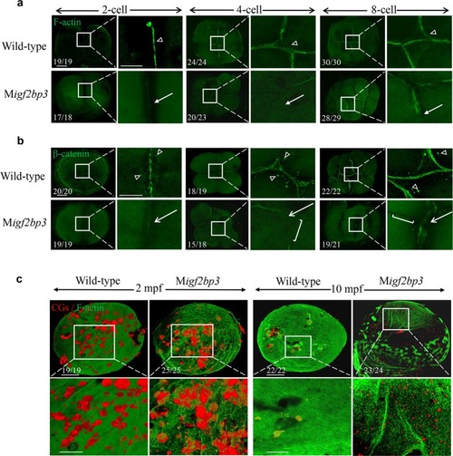
M<italic>igf2bp3</italic> embryos exhibit abnormal cytoskeleton organization.a, b Expression and distribution of F-actin and β-catenin in wild type and Migf2bp3 embryos at 2-, 4-, and 8-cell stages. The F-actin and β-catenin were enriched in mature and apparent cleavage furrow (triangle). The inset showed a zoom-in of the boxed region at each stage. Zoomed-out scale bar = 100 μm; zoomed-in scale bar = 50 μm. a Migf2bp3 embryos at 4-cell stage did neither exhibit intact adhesion junction of F-actin cables nor form the mature furrow (white arrow). b Migf2bp3 embryos exhibited reduced expression of β-catenin (white arrows) and did not form intact cell adhesion junction with defective furrow (white bracket). c Double localization of F-actin (green fluorescence) and CG (red fluorescence). The inset showed a zoom-in of the boxed region at each stage. Zoomed-out scale bar = 100 μm; zoomed-in scale bar = 50 μm.
|