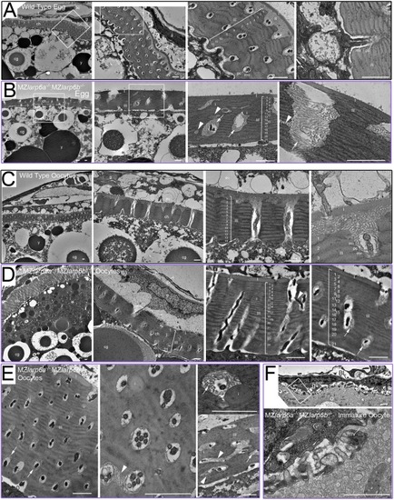
Larp6 maternal effect on oocyte development and chorion structure. (A-F) Transmission electron micrographs of mature unactivated eggs (A,B) and ovaries (C-F) from wild-type (A,C) and mutant (B,D-F) MZlarp6a−/−;MZlarp6b−/− mothers. (A) Unactivated wild-type eggs contain yolk platelets (y) and cortical granules (cg), are closely surrounded by a tri-zoned chorion (ch), and are beneath the remains of supporting granulosa (g) and thecal (t) cells separated by basal lamina (BL). There is a smooth thin outer chorionic zone (I), a fibrillar meshwork second zone (II) and a multi-layered thick inner zone (III) consisting of 12-14 alternating dark and light sublayers punctured by regularly spaced transverse pore canals (pc) that sometimes contain microvilli (mv): cellular processes that are ∼100 nm in diameter. (B) Eggs from a mutant mother contain: similar yolk platelets and cortical granules; gross defects in chorionic structure, with irregular pores (arrowheads) and inclusions (i); increased numbers (17-30) of thinner sub-layers in zone III; no zone II; mispositioning of zone II fibrillar material deep into pores (arrows); and an apparently normal zone I. (C) Wild-type ovarian tissue contains eggs showing a series of developmental stages of chorion development. Large oocytes have a 12-14 layered zone III that retain bidirectional processes in regular pores. Zone II fibrillar material is not present deep within the pores. (D,E) Ovarian tissue from a larp6a−/−;larp6b−/− mutant female shows defective chorion structure in several separate oocytes (D) and, at high magnification, mutant oocytes have irregular and branching pores (arrowheads), up to six process profiles per pore canal, disorganised sub-layering in zone III (numbers), absence of a uniform zone II layer, parallel bundling of zone II fibrils and penetration of fibrillar material through the entire depth of pores (arrows) (E). (F) An immature double mutant oocyte lacking yolk platelets and cortical granules had granulosa cells closely opposed to the oocyte plasma membrane with some regions of interdigitating processes initiating chorionogenesis that were indistinguishable from wild type. Boxes show successively magnified areas in A,B (first three panels only) and F. Scale bars: 1 μm.
|
