
Fig. 4
- ID
- ZDB-FIG-200313-7
- Publication
- Karjosukarso et al., 2020 - Modeling ZNF408-Associated FEVR in Zebrafish Results in Abnormal Retinal Vasculature
- Other Figures
- All Figure Page
- Back to All Figure Page
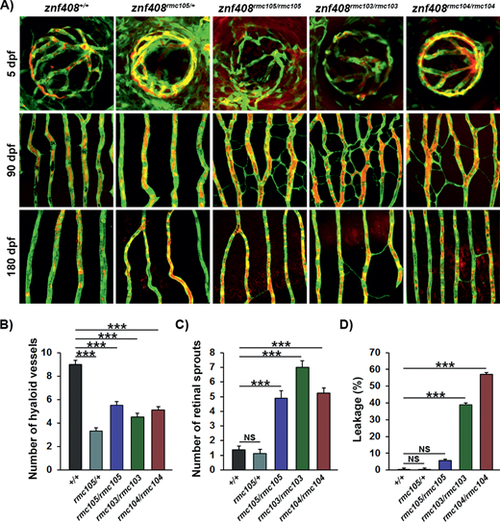
Progressive vascular pathology in znf408-mutant zebrafish larvae and adults. (a) Confocal micrographs at 5, 90, or 180 days postfertilization (dpf) of the hyaloid (5 dpf, top row) or retinal vessels (90 dpf, second row and 180 dpf, third row) in znf408+/+, znf408rmc105/+, znf408rmc105/rmc105, znf408rmc103/rmc103, and znf408rmc104/rmc104 zebrafish crossed onto the Tg(flk1:GFP) background (vessels shown in green) following injection with rhodamine-conjugated dextran (plasma shown in red). (b) Quantification of the number of hyaloid vessels at 5 dpf from the groups shown in (a). n = 10, ANOVA: P < 0.001, Dunnett's post hoc: ***P < 0.001. (c) Quantification of the number of sprouts in the retinal vessels at 90 dpf from the groups shown in (a). n = 10, ANOVA: P < 0.001, Dunnett's post hoc: ***P < 0.001. (d) Quantification of the leaked dextran as a percentage of total dextran in the retinal vasculature at 180 dpf from the groups shown in (a). n = 10, ANOVA: P < 0.001, Dunnett's post hoc: ***P < 0.001. |
| Fish: | |
|---|---|
| Observed In: | |
| Stage Range: | Day 5 to Adult |