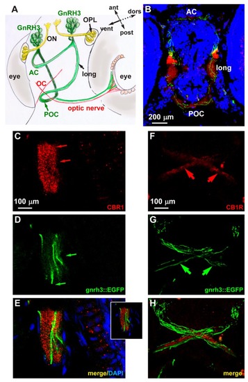
Expression of CB1R in developing zebrafish brain. (A) Scheme illustrating the position of the GnRH3 neurons relative to the position and orientation of the main forebrain commissure in the zebrafish brain (redrawn from [52]). (B) Low magnification double-fluorescent images of GnRH3::EGFP neurons (green) and IFL with an anti-CB1R antibody (red) on cryostatic sections of the zebrafish head at the age 72 hpf. (C–E) Higher magnification double-fluorescence images of the anterior commissure, showing overlapping localization of CB1R punctate staining (red) with EGFP+ fibers (green). Sections were counterstained with DAPI (blue). The merged signal is shown in E. Inset in E shows a single stack of the same image. Arrows indicate the fluorescence signal. (F–H) Higher magnification double-fluorescence images of the optic chiasm, immunostained for CB1R (red), as in panels C–E. Adjacent but non-overlapping expression was observed in these fibers. Arrows indicate the fluorescence signal. Scale bars are reported in panels B, in C (for C,D) and in F (for F–H). AC, anterior commissure; ant, anterior; dors, dorsal; long, longitudinal tract; OC, optic chiasm; ON, olfactory nerve; OPL, olfactory placode; POC, postoptic commissure; post, posterior; RGC, retinal ganglion cells; ven, ventral.
|
