|
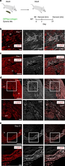
Adult mouse splenic GFP<sup>+</sup> monocytes contribute collagen to the scar post-MI.a Experimental design for adoptive transfer: monocytes were isolated by magnetic column purification from the spleens of adult GFPtpz-collagen+ mouse donors. At the time of MI surgery, 50 ± 10 × 103 monocytes/PBS, or PBS alone, were transferred to wild type (WT) recipients by intracardiac injection. Adult recipient mice were then harvested at either day 7 (b, c) or day 21 (d, e) for immunostaining. b At day 7 post-MI, combined immunostaining for α-GFP (red; to exclude autofluorescence from monitoring GFP-alone) and α-CD68 revealed GFP+ macrophages within the scar region (sc) of the left ventricle (lv). c Co-staining for collagen-1 (white, Col1) revealed GFP+ collagen deposits within the same scar region. d By day 21, there was increased GFP+ collagen deposition within the scar (white inset boxes in upper panels shown at higher magnification in corresponding panels below). e GFP+ collagen fibres were evident within regions of transmural scar, as detected by combined α-GFP and Col1 staining (white inset boxes in upper panels shown at higher magnification in corresponding panels below). lv, left ventricle, pm, papillary muscle; sc, scar. Scale bars: b, c upper panel, d upper panel 100 μm; c lower panel, d lower panel 50 μm. Representative images of n = 3 per group.
|