FIGURE

Figure 5

ID
ZDB-FIG-200129-38
Publication
Fallata et al., 2019 - Intracellular Localization in Zebrafish Muscle and Conserved Sequence Features Suggest Roles for Gelatinase A Moonlighting in Sarcomere Maintenance
Other Figures
All Figure Page
Back to All Figure Page
Figure 5

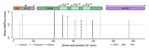
Gelatinase A orthologues have highly conserved phosphorylation sites. Putative serine (solid lines), threonine (dashed lines), and tyrosine (dotted lines) phosphorylation sites conserved in 100% (black), 99% (dark grey), or 97% (light grey) of gelatinase A orthologues are shown with respect to a structural schematic of the gelatinase A protein, illustrating the signal sequence (1–29 (orange)), propeptide (30–107 (grey)), catalytic domain (118–446 (green)) with fibronectin-like repeats (light green), and hemopexin-like domain (463–657 (purple)). Cysteines are indicated with yellow spots, connected by horizontal lines if they are predicted to participate in intramolecular disulfide bonds. Conserved residues that have been empirically demonstrated to be phosphorylated in vivo in the human protein are indicated with asterisks.

Expression Data

Expression Detail
Antibody Labeling
Phenotype Data

Phenotype Detail
Acknowledgments
This image is the copyrighted work of the attributed author or publisher, and ZFIN has permission only to display this image to its users. Additional permissions should be obtained from the applicable author or publisher of the image. Full text @ Biomedicines