FIGURE

FIGURE 2

ID
ZDB-FIG-200112-19
Publication
Rosch et al., 2019 - Functional Genomics of Epilepsy and Associated Neurodevelopmental Disorders Using Simple Animal Models: From Genes, Molecules to Brain Networks
Other Figures
All Figure Page
Back to All Figure Page
FIGURE 2

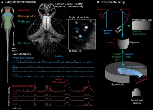
Recording whole-brain dynamics at single-cell resolution in zebrafish models of neurodevelopmental disorders. (A) Larval zebrafish at 7 days post fertilization are freely behaving and have all the major anatomical subdivisions of the vertebrate brain (left). Transgenic lines expressing genetically encoded calcium indicators in neurons can be used to record neuronal function through fluorescence signals. Because of their small size, the whole brain can be captured at single cell resolution (top). This allows recording of whole brain dynamics alongside single-cell behavior (bottom). (B) Zebrafish larvae can be embedded in transparent agarose, allowing in vivo imaging using fluorescence microscopy (shown here is a two-photon microscopy setup). Depending on the experimental paradigm, behavioral output can further be tracked using recordings of tail movements in tail free set ups. This allows e.g., linking of convulsive movements and brain hypersynchrony to identify epileptic seizures in the zebrafish.

Expression Data

Expression Detail
Antibody Labeling
Phenotype Data

Phenotype Detail
Acknowledgments
This image is the copyrighted work of the attributed author or publisher, and ZFIN has permission only to display this image to its users. Additional permissions should be obtained from the applicable author or publisher of the image. Full text @ Front. Cell. Neurosci.