FIGURE

Fig. 4

ID
ZDB-FIG-200103-4
Publication
Zhang et al., 2019 - Expression analysis of Rab11 during zebrafish embryonic development
Other Figures
All Figure Page
Back to All Figure Page
Fig. 4

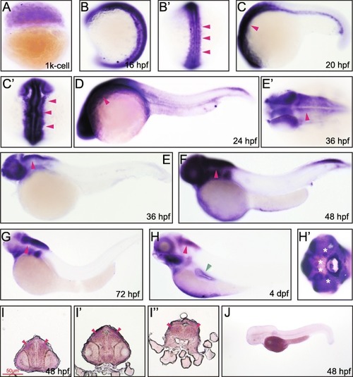
Whole mount in situ and transverse section hybridization analysis of rab11ba in zebrafish embryos. A 1 k cell, lateral view. B 16 hpf, lateral view. B’ 16 hpf, dorsal view, arrowheads indicate neural tube. C 20 hpf, lateral view, arrowhead indicates brain. C’ 20 hpf, dorsal view, arrowheads indicate brain. D 24 hpf, lateral view, arrowhead indicate brain. E 36 hpf, lateral view, arrowhead indicates spinal cord. E’ 36 hpf, dorsal view, arrowhead indicates brain and head structure. F 48 hpf, lateral view, arrowhead indicates spinal cord. G 72 hpf, lateral view, arrowhead indicates brain. H 4 dpf, lateral view, red arrowhead indicates brain, green arrowhead indicates pectoral fins. H’ 4 dpf, asterisks indicate neuromasts. I 48 hpf, the trunk transverse section of the embryonic forebrain, arrowheads indicate cerebral cortex. I’ 48 hpf, the trunk transverse section of the embryonic midbrain, arrowheads indicate cerebral cortex. I” 48 hpf, the trunk transverse section of the embryonic hindbrain, arrowheads indicate cerebral cortex. J 48 hpf, lateral view, embryos stained with the sense rab11ba probe

Expression Data
Gene:
Fish:
Anatomical Terms:
Stage Range: 1k-cell to Day 4

Expression Detail
Antibody Labeling
Phenotype Data

Phenotype Detail
Acknowledgments
This image is the copyrighted work of the attributed author or publisher, and ZFIN has permission only to display this image to its users. Additional permissions should be obtained from the applicable author or publisher of the image. Full text @ BMC Dev. Biol.