FIGURE

Fig. 6

ID
ZDB-FIG-191230-672
Publication
Pérez-Rius et al., 2019 - Comparison of zebrafish and mice knockouts for Megalencephalic Leukoencephalopathy proteins indicates that GlialCAM/MLC1 forms a functional unit
Other Figures
All Figure Page
Back to All Figure Page
Fig. 6

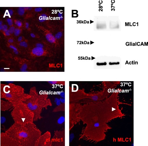
zfmlc1 and hMLC1 overexpressed in primary Glialcam−/− astrocytes are located at cell-cell junctions. a, b Primary astrocytes isolated from Glialcam−/− mice were incubated for 18 h at 28 °C. Then, MLC1 was detected by immunofluoresce (a) and protein levels were monitored by Western blot (b). Actin served as a loading control. Lack of signal using GlialCAM antibodies confirmed the lack of protein expression. c, d Overexpression using adenoviruses of mlc1 from zebrafish (zfmlc1, c) and human HA-tagged MLC1 (hMLC1, d) detected both MLC1 proteins at cell-cell junctions (arrowheads) in primary astrocytes isolated form Glialcam−/− mice. We used antibodies detecting the zebrafish MLC1 or the HA epitope, which did not detect the endogenous Mlc1. Scale bar: 10 μm

Expression Data

Expression Detail
Antibody Labeling
Phenotype Data

Phenotype Detail
Acknowledgments
This image is the copyrighted work of the attributed author or publisher, and ZFIN has permission only to display this image to its users. Additional permissions should be obtained from the applicable author or publisher of the image. Full text @ Orphanet J Rare Dis