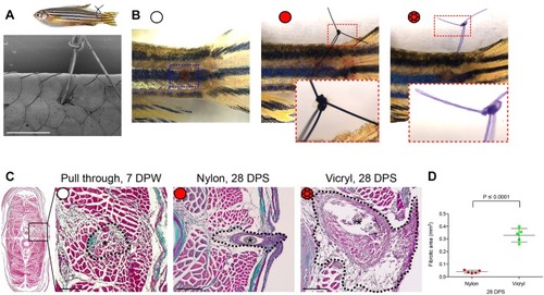
Extent of foreign body fibrotic encapsulation is dependent on suture type. (A) Schematic illustration of the zebrafish suture model, with scanning electron micrograph showing a suture in place. n=6 independent fish. (B) Representative images of zebrafish following suture pull through (white circle), nylon suture (red circle) or vicryl suture (red braided circle), at 1 day post suturing (DPS) with insets to show suture detail. n=5 independent fish per condition. (C) Masson's Trichrome-stained transverse sections of pull through at 7 DPW, and nylon or vicryl sutured fish at 28 DPS, to indicate extent of fibrosis. Sutures are indicated with black asterisks; the zone of scarring and fibrotic encapsulation is indicated by black dotted line overlay. n=5 independent fish per condition. (D) Quantification of total area of fibrotic encapsulation, measured from images in C. Error bars in D indicate mean±s.d. Statistical significance is indicated, as determined by two-tailed t-test. Scale bars: 1 mm (A,B), 100 μm (C).
|
