|
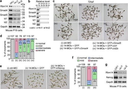
The ventralization defects of <italic>rbm14</italic> morphants is mainly attributed to insufficient BMP signaling.a, b Knockdown of mRbm14 in P19 cells concomitantly downregulated Smad4, Smad5, Id1, and Id2. P19 cells transfected with control siRNA (ctrl-i) or each of the two mRbm14-specific siRNAs (14-i1 and 14-i2) for 48 h were collected and subjected to immunoblotting. Gapdh served as a loading control. The quantification results (b), presented as mean ± SD, were based on band intensities from two independent experiments. c, d Overexpressing smad4, smad5, id1, or id2b attenuated the ventralization defects of rbm14 morphants. Zebrafish embryos at the one-cell stage were co-injected with the indicated MOs (total 8 ng per embryo) and in vitro-transcribed mRNA (300 pg per embryo) coding for GFP or the GFP-tagged proteins (also see Supplementary Fig. 2). Those injected with ctrl-MO served as a negative control. e, f Overexpressing Nanog attenuates the ventralization defect of zebrafish rbm14 morphants. Zebrafish embryos at the one-cell stage were co-injected with the indicated MOs (total 8 ng per embryo) and in vitro-transcribed mRNA coding for GFP or GFP-mNanog (300 pg per embryo) (also see Supplementary Fig. 2). Embryos injected with ctrl-MO served as a negative control. Quantification results (d, f), based on the criteria and examples in Fig. 1e and presented as mean ± SD, were from three independent experiments. Student’s t-test against the GFP mRNA-injected populations: n.s., no significance (P > 0.05); *P < 0.05; **P < 0.01; ***P < 0.001. Total number of embryos analyzed are listed over each histogram. g Depletion of Rbm14 in P19 cells downregulated Nanog. P19 cells transfected with the indicated siRNAs for 48 h were subjected to immunoblotting. Gapdh served as a loading control
|